At TheHealthBoard, we're committed to delivering accurate, trustworthy information. Our expert-authored content is rigorously fact-checked and sourced from credible authorities. Discover how we uphold the highest standards in providing you with reliable knowledge.
What Is Contrast Echocardiography?
Contrast echocardiography is a type of echocardiogram, which is a cardiac ultrasound and can also be called an echo. By adding a contrast, a cardiac echo has better resolution. The additional, clearer resolution allows doctors to watch blood flow through the heart. Contrast is most commonly a saline solution, although research continues to find other sources for contrast.
An echocardiogram is a special type of ultrasound. It is used specifically in cardiac applications to provide images of the heart. Standard echocardiographyuses Doppler sound waves to provide images of tissue and blood velocity. Contrast echocardiography adds a contrast for different cardiac images. The contrast provides extra visibility so that enhanced imaging of tissue and blood flow is possible.

Saline solution is used in contrast echocardiography. The saline is gently agitated to create microbubbles and is then injected into a vein. During the cardiac echo, the bubbles reflect sound waves, which make them appear opaque. Unlike the red blood cells, these bubbles allow doctors to monitor the volume and the exact flow of blood in and out of the heart.

The patient scheduled for contrast echocardiography will be instructed to remove clothing from the waist up. Special gel used for an ultrasound is applied to the skin around the heart area, and a standard echo is performed for basic imaging. An intravenous (IV) line is then inserted into the vein on the left arm above the elbow. This IV line will provide access for the sonographer to inject contrast as needed. Once the contrast is agitated, it must be immediately injected because the bubbles are delicate and do not have a long amount of time before they disappear.

Contrast echocardiography provides doctors with the ability to see the functions of the heart and blood flow. Errors in heart function or problems with blood flow can be visible by adding contrast to a standard echocardiogram. The procedure is non-invasive and does not require surgery or scopes. A cardiac ultrasound is performed on the surface of the body.
Doctors use contrast echocardiography to diagnose a variety of heart issues. Myocardial echocardiography, for example, uses contrast to gain images of the myocardial capillary vascular bed in the heart. A stress echocardiogram uses contrast to monitor systolic function and how ventricular walls are moving during activity. Both of these examples allow doctors to see problems with the heart that may not be otherwise noticed, particularly with a standard echocardiogram.
AS FEATURED ON:
AS FEATURED ON:

-
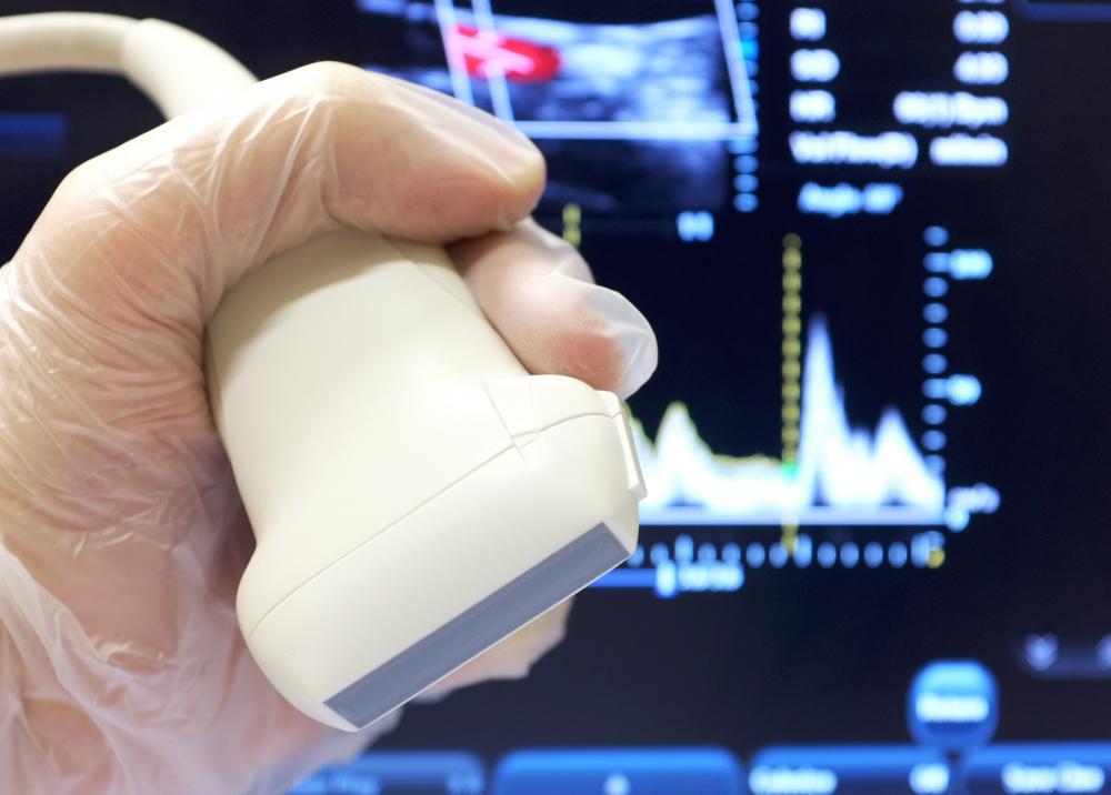
![An echocardiogram is a specialized type of ultrasound that only documents the cardiac system.]() By: zayacskAn echocardiogram is a specialized type of ultrasound that only documents the cardiac system.
By: zayacskAn echocardiogram is a specialized type of ultrasound that only documents the cardiac system. -
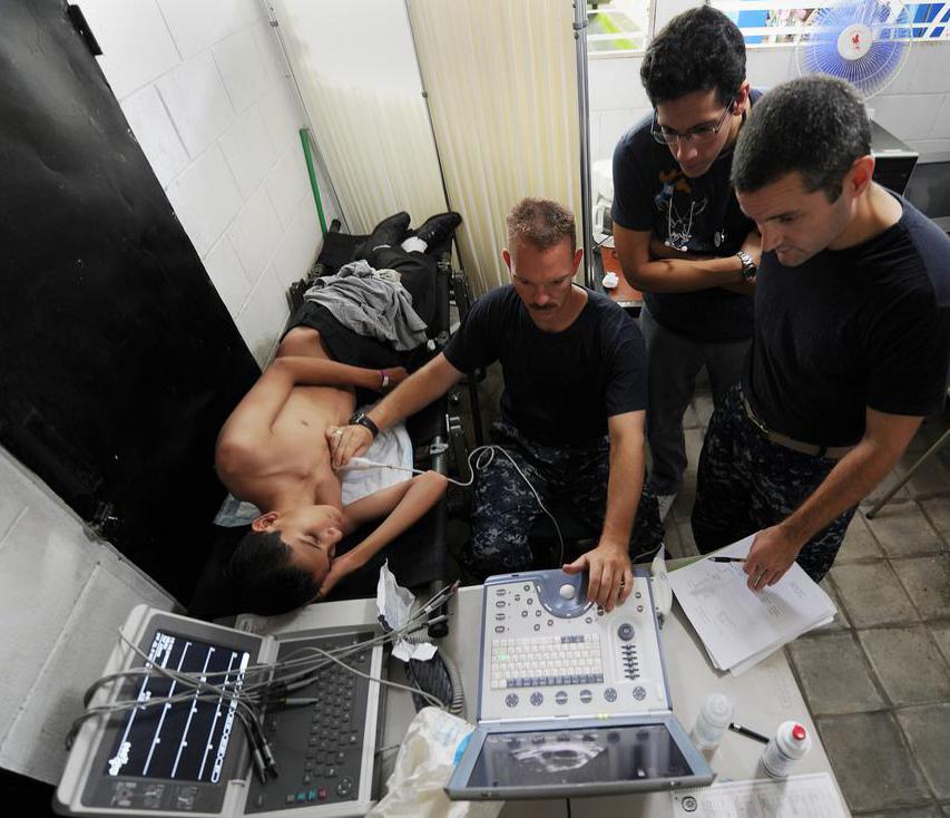
![An echocardiogram is typically performed by an ultrasound technician.]()
An echocardiogram is typically performed by an ultrasound technician. -
![Doctors use contrast echocardiography to diagnose a variety of heart issues.]() By: lightwavemediaDoctors use contrast echocardiography to diagnose a variety of heart issues.
By: lightwavemediaDoctors use contrast echocardiography to diagnose a variety of heart issues.

Discuss this Article
Post your comments