At TheHealthBoard, we're committed to delivering accurate, trustworthy information. Our expert-authored content is rigorously fact-checked and sourced from credible authorities. Discover how we uphold the highest standards in providing you with reliable knowledge.
What Is the Treatment for Macular Edema?
Treatment for macular edema may involve medications, laser therapy, surgery, or a combination of methods depending on the cause and the seriousness of the condition. The edema may occur secondary to aging, existing medical conditions, or trauma. The disorder typically involves increased pressure on or near the macula portion of the retina.
Physicians typically determine treatment for macular edema based on ocular testing that indicates whether the problem lies in swollen tissue or blood vessel leakage. Optical coherence tomography, or OCT, involves visualizing the interior of the eye for indications of macular swelling or thickening. Fluorescein angiography provides intraocular photographs taken with a special camera that emits blue flashes. Patients generally receive an intravenous dye prior to the photographic study. The dye illuminates blood vessels and possible points of hemorrhage.

When uveitis, or intraocular inflammation, leads to generalized edema, physicians may prescribe anti-inflammatory or steroidal eye drops. When inflammation occurs secondary to diabetic retinopathy, patients may require oral steroids or intraocular steroid injections. Treatment for macular edema caused by glaucoma usually involves eye drops appropriate for closed angle or open angle varieties of the disorder.

Biochemical changes that accompany age-associated macular degeneration, diabetic retinopathy, or genetically acquired retinitis pigmentosa may increase production of vascular endothelial growth factor, or VEGF, which promotes blood vessel development. Physicians may prescribe medications that prevent VEGF release to curtail further vessel development.

The blood vessels formed under these circumstances are usually fragile and may hemorrhage. The bleeding that occurs depletes necessary blood and oxygen while distorting the light rays that enter the eye. Specialists use vitrectomy, sometimes combined with laser surgery, as treatment for macular edema caused by hemorrhage. Surgeons may block or remove immature blood vessels, blood and protein plaques to prevent further bleeding and clarify vision.

The macula comprises a minute portion of the retina located at the back of the eye. This region contains nerve cells, known as cones, that are responsible for visualizing color and for general sight. When the macula swells, the increased pressure may decrease the blood and oxygen supply, distorting vision. These areas may also endure compression from increased fluid in the vitreous humor. Either scenario indicates macular edema.

Symptoms of edema generally affect areas either to one side or to a central region within the visual field. These areas generally appear blurry or washed out. Individuals may feel as if they are gazing through a semi-opaque plastic film. Some patients may experience an overall pinkish cast in the affected eye. Left untreated, macular edema usually progresses and may damage vision permanently.
AS FEATURED ON:
AS FEATURED ON:

-
![Laser surgery may be used as part of a treatment for macular edema.]() By: Monkey BusinessLaser surgery may be used as part of a treatment for macular edema.
By: Monkey BusinessLaser surgery may be used as part of a treatment for macular edema. -
![Macular edema can be caused by glaucoma.]() By: joshyaMacular edema can be caused by glaucoma.
By: joshyaMacular edema can be caused by glaucoma. -
![A macular edema may occur secondary to aging.]() By: ChmpagnDaveA macular edema may occur secondary to aging.
By: ChmpagnDaveA macular edema may occur secondary to aging. -
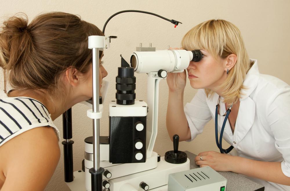
![Ocular testing can be used to investigate the cause of macular swelling.]() By: JackFOcular testing can be used to investigate the cause of macular swelling.
By: JackFOcular testing can be used to investigate the cause of macular swelling. -
![If macular edema remains untreated, the disorder can cause blurry or otherwise distorted vision.]() By: bellemediaIf macular edema remains untreated, the disorder can cause blurry or otherwise distorted vision.
By: bellemediaIf macular edema remains untreated, the disorder can cause blurry or otherwise distorted vision. -
![It's important to have periodic eye exams, particularly in cases of vision changes or abnormalities.]() By: FotolEdharIt's important to have periodic eye exams, particularly in cases of vision changes or abnormalities.
By: FotolEdharIt's important to have periodic eye exams, particularly in cases of vision changes or abnormalities. -
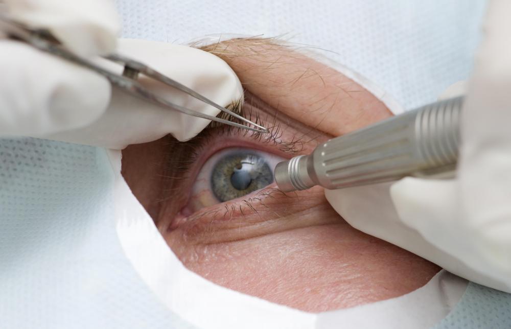
![Treatment for macular edema may include eye surgery.]() By: Max TacticTreatment for macular edema may include eye surgery.
By: Max TacticTreatment for macular edema may include eye surgery.


Discuss this Article
Post your comments