At TheHealthBoard, we're committed to delivering accurate, trustworthy information. Our expert-authored content is rigorously fact-checked and sourced from credible authorities. Discover how we uphold the highest standards in providing you with reliable knowledge.
What is a Cavernous Sinus Meningioma?
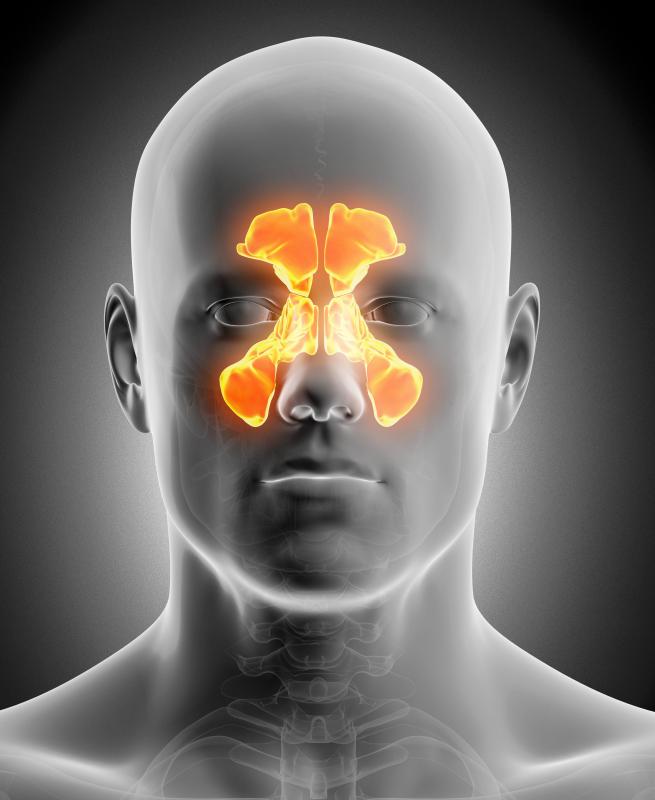
The paired cavernous sinuses are thin-walled venous pockets below and behind the orbits or eye sockets that collect blood draining from the face, eye, and anterior brain. A cavernous sinus meningioma is a benign tumor arising from the cells that form the internal lining membrane of the brain, called the pia mater, which expands to fill the cavernous sinus. The cavernous sinus has many vital structures passing through it, including the carotid artery and the third, fourth, fifth, and sixth cranial nerves. Most of the predominant symptoms of a cavernous sinus meningioma are created by compression of these structures by the expanding tumor, producing cavernous sinus syndrome. These symptoms include pain behind the eye, ocular redness, blurred or double vision, difficulty with eye movement, and forward bulging of the eye.
A patient with a cavernous sinus meningioma may suffer palsies of the third, fourth, and sixth cranial nerves, producing varying degrees of impairment with eye movement. Typically, the patient has pain with attempts at eye movement. The decreased venous drainage from the eye dilates the surface veins on the eye, giving the eye a reddish hue. The eye pressure may also be elevated, and the optic disc may appear to be swollen or pale, with surrounding hemorrhages. Furthermore, the pupil may be fixed in the mid-dilated position and non-reactive, and the patient may be numb to sensation on the eye surface and the face.

When physicians suspect a cavernous sinus meningioma, the patients should undergo multiplanar scans with magnetic resonance imaging (MRI) or computed tomography (CT) of the orbits and cavernous sinus regions. Although CT scans offer enhanced visualization of the bones, MRI scans provide better resolution of the soft-tissue structures of these regions. Physicians often order contrast dye to accent the blood-filled structures, such as the superior and inferior orbital veins that drain into the cavernous sinus and the internal carotid artery that travels through the sinus. Pre-contrast and post-contrast scans provide useful information, with the tumors isointense on T1-weighted MRIs with dense enhancement after injection of the contrast. CT scans show an associated thickening and hypercalcification of the adjacent bone.

A patient with a cavernous sinus meningioma has three basic treatment options. Some physicians recommend observation alone for those patients in whom the tumors are growing slowly and producing few symptoms. Patients with serial growth or progressively worsening symptoms may need to undergo microsurgical removal of the tumor through the skull base with a variety of approaches. Some treatment centers use a gamma knife and linear accelerator to remove the tumor radiologically. In all forms of surgical management, surgeons must attempt to remove the entire tumor while sparing the internal carotid artery.
AS FEATURED ON:
AS FEATURED ON:

-
![A linear accelerator might be used to target a cavernous sinus meningioma with radiation.]() By: Alex TihonovA linear accelerator might be used to target a cavernous sinus meningioma with radiation.
By: Alex TihonovA linear accelerator might be used to target a cavernous sinus meningioma with radiation. -
![A patient with a cavernous sinus meningioma may have pupils that are fixed in the mid-dilated position.]() By: ra2 studioA patient with a cavernous sinus meningioma may have pupils that are fixed in the mid-dilated position.
By: ra2 studioA patient with a cavernous sinus meningioma may have pupils that are fixed in the mid-dilated position. -
![An MRI scan will provide great resolution of the soft-tissue structures of the cavernous sinus regions.]() By: James SteidlAn MRI scan will provide great resolution of the soft-tissue structures of the cavernous sinus regions.
By: James SteidlAn MRI scan will provide great resolution of the soft-tissue structures of the cavernous sinus regions. -
![The cavernous sinuses are located below and behind the eye sockets.]() By: Sebastian KaulitzkiThe cavernous sinuses are located below and behind the eye sockets.
By: Sebastian KaulitzkiThe cavernous sinuses are located below and behind the eye sockets. -
![An individual with a cavernous sinus meningioma may notice blurred or double vision.]() By: bellemediaAn individual with a cavernous sinus meningioma may notice blurred or double vision.
By: bellemediaAn individual with a cavernous sinus meningioma may notice blurred or double vision. -
![A cavernous sinus meningioma can cause pain behind the eye, as well as abnormal eye movement.]() By: FotolEdharA cavernous sinus meningioma can cause pain behind the eye, as well as abnormal eye movement.
By: FotolEdharA cavernous sinus meningioma can cause pain behind the eye, as well as abnormal eye movement. -
![Gamma knife radiosurgery is one of the most precise and effective treatments available for a cavernous sinus meningioma.]() By: Monkey BusinessGamma knife radiosurgery is one of the most precise and effective treatments available for a cavernous sinus meningioma.
By: Monkey BusinessGamma knife radiosurgery is one of the most precise and effective treatments available for a cavernous sinus meningioma.


Discuss this Article
Post your comments